Necrotizing Fasciiti


Necrotizing fasciitis is infection of skin and fascia caused by group A streptococci, mixed aerobic-anaerobic bacteria, or Clostridium perfringens. It develops rapidly and has high mortality without emergent treatment.

Signs and Symptoms:
  • Pain and unexplained fever
  • Swelling, tenderness, induration, or bullae
  • Extending of infection to fascia and spreads rapidly
  • Predisposing factors: DM, peripheral vascular disease, breaches in the skin or mucosa, and surgery
  • Necrotizing fasciitis of the perineal region is called Fournier's gangrene
Treatments:
  1. Surgery
  2. Antibiotic therapy after gram stain and culture of tissue

Molluscum contagiosum


Molluscum contagiosum is an infection caused by a poxvirus. It is commonly found in young children and AIDS patients

Signs and Symptoms:
  • Discrete, dome shaped, and shiny papules (2-5 mm) with central umbilication
  • The lesions are asymptomatic unless inflamed or irritated
  • In adult: lesions on the perianal and perigenital areas
  • In child: lesions on the trunk, extremities, and face
Treatments:
  1. The lesions resolve (months to years) spontaneously
  2. Liquid nitrogen cryotherapy
  3. Curettage
  4. Trichloroacetic acid

Lice


Types of Lice:
  1. Pediculus capitis - infection of the scalp
  2. Pediculus corporis - infection of the body
  3. Phthirus pubis - infection of the pubic area
Signs and Symptoms:
  • Pruritus
  • Nits fluoresce blue under Wood's lamp
Treatments:
  • Permethrin shampoo or cream
  • Sources of reinfection (such as combs, bed sheets, clothing) should be decontaminated

Lyme Disease


Lyme Disease:
  • "Bull's eye" erytheuma
  • Occurs in moist and warm areas of the body (axillae, behind the knees, inguinal area) where the tick initially bites
  • Fatigue
  • Malaise
  • Arthralgias
  • Headache
  • Lymphadenopathy
  • Neurologic deficits
  • History of tick bite

Rocky Mountain Spotted Fever


Rocky Mountain Spotted Fever:
  • Rash on the palms and soles then spreads to the trunk and face
  • High fever
  • Chills
  • Headache
  • History of tick bite

Scarlet Fever


Scarlet Fever:
  • Rash on the trunk as sandpaper-like
  • Strawberry tongue
  • Circumoral pallor
  • Most occurs in untreated streptococcal pharyngitis
  • Penicillin is drug of choice to prevent rheumatic fever

Chickenpox (varicella)


Chickenpox (varicella):
  • Macules on the trunk cause papules and vesicles, ruptured and crusting
  • Crops of lesions
  • Dewdrop on a rose petal: a clear vesicle on an erythematous base
  • Fever
  • Risk for bacterial superinfection of lesions

Fifth Disease


Fifth Disease (Erythema infectiosum):
  • Caused by parvovirus B19
  • Slapped-cheek rash over the cheeks
  • Maculopapular rash on the trunk and extremities 1-2 days later
  • Malaise
  • Low-grade fever

Infectious Rashes: Rubella


Rubella (German measles):
  • Maculopapular rash (spreading from the head downward)
  • Suboccipital swelling
  • Postauricular nodes
  • Low-grade fever
  • Malaise
  • Arthralgias

Infectious Rashes: Measles / Rubeola


Measles / Rubeola:
  • Koplik's spots (white spots on buccal mucosa) usually occurs 3 days after a high fever
  • Maculopapular rash (spreading from the head downward)
  • Cough
  • Coryza
  • Conjunctivitis

Impetigo


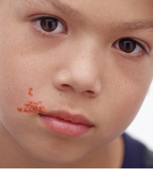
Impetigo is a contagious and autoinoculable skin infection caused by staphylococci or streptococci. It commonly in children than adults

Signs and Symptoms:
  • Pruritus with honey-colored crusts on the face, neck, and extremities
  • Nonbullous impetigo: superficial pustules with surrounding erythema
  • Bullous impetigo: erythematous macules as thin-walled vesicles or bullae on an erythematous base.
Treatments:
  1. Mild soap for washing the lesions
  2. Topical mupirocin
  3. Systemic antibiotics
  4. Patient's towels and washcloths should be segregated

Herpes Simplex


Herpes simplex is a recurrent vesicular eruption of mucocutaneous surfaces due to HSV infections. It is a painful eruption.

Signs and Symptoms:
  • First outbreaks are longer and more sever than recurrent eruptions
  • Prodromal tingling, burning, or frank pain with lymphadenopathy
  • Fever, discomfort, malaise, and edema on involved area
  • Grouped vesicles on erythematous base
  • Recurrent infections are limited to mucocutaneous areas
Treatments:
  1. Acyclovir ointments
  2. Oral or IV acyclovir
  3. Daily acyclovir therapy for patient with more than 6 outbreaks per year

Fungal Infections: Tinea Corporis/Capitis


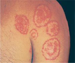
Tinea corporis / capitis is an infection of the body/scalp caused by fungus

Signs and Symptoms:
  • Pruritus
  • Ring-shaped (ringworm)
  • Erythematous
  • Scaling plaques
Treatments:
  1. Tinea corporis: topical anti-fungal
  2. Tinea capitis and tinea unguium (nail infection): oral anti-fungals

Fungal Infections: Pityriasis versicolor


Pityriasis versicolor is also named by tinea versicolor. It is caused by Malassezia furfur.

Signs and Symptoms:
  • Small, scaling, hyper or hypopigmented macules
  • Lesions: asymptomatic or mild itching
  • Chest and back is the common site of infections
Diagnosed by:
Treatments:
  1. Topical anti-fungal agents
  2. Selenium sulfide shampoo

Cellulitis


Cellulitis or folliculitis is skin infections due to streptococci or staphylococci.

Signs and Symptoms:
  • Folliculitis: hair follicles infection commonly on facial hair
  • Cellulitis: red, hot, and swollen skin lesions, fever and chills
Treatments:
  1. Oral antibiotics such as cephalexin or dicloxacillin for mild to moderate cases
  2. In cases of systemic toxicity, comorbid condition, DM, extremes of age, or hand or orbital involvement, patient should be hospitalized and give IV antibiotics

Acne Vulgaris


Acne vulgaris is a condition in which the hair follicles are blocked by sebum and keratinous material. This blockage causes formation of cyst/comedones. Propionibacterium acnes within comedones break down sebum into free fatty acids.

History and Physical Exams:
  • Acne is associated with changes in androgen levels
  • Found primarily in puberty
  • More common in males than in females
  • Open comedones - blackheads and closed comedones - whiteheads on the face, neck, arms, back, and buttocks
  • Drugs are related to acne: lithium, steroids, OCPs, androgens
Treatments:

  1. Treatments are according to the severity of acne
  2. Mild case: topical clindamycin or erythromycin, benzoyl peroxide, topical retinoids
  3. Moderate case: treatments in mild case plus oral antibiotics
  4. Severe case: oral isotretinoin (Accutane)

Vitiligo


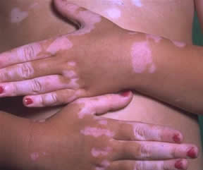
Vitiligo is a depigmentation due to loss of melanocytes and associates with autoimmune and thyroid disease. It more common in dark-skinned peaople with a family history.

Signs and Symptoms:
Irregular white patches

Treatments:

Steven-Johnson Syndrome


Steven-Johnson Syndrome/Toxic Epidermal necrolysis (SJS/TEN) is a life threatening exfoliative mucocutaneous disease. It often caused by a drug induce immunologic reaction such as sulfa drugs, carbamazepine, phenytoin, valproic acid, quinolones, cephalosporins, phenobarbital, allopurinol, aminopenicillins, and corticosteroids.

Signs and Symptoms:
  • History of new drug exposure within 1-3 weeks
  • Flu-like prodrome
  • Skin tenderness
  • Painful mouth lesions
  • Nikolsky's sign is positive
  • Coalescing red macules and flaccid blisters with full-thickness epidermal loss
  • Sepsis from superimposed bacterial skin infections that leads to death
Treatments:

  1. Eliminate the offending agents
  2. Analgesia and IVIG
  3. Hospitalize in the burn center to manage skin and fluid losses

Seborrheic Dermatitis


Seborrheic dermatitis is a chronic superficial inflammatory. It may be a reaction to Pityrosporum yeast

Signs and Symptoms:
  • Pruritus during neonatal and postpubertal periods
  • Yellowish, greasy, and erythematous scaling patches and plaques on the scalp, ear, and face
Treatments:
  • 1% hydrocortisone and topical antifungals
  • Shampoos contain of selenium sulfide or zinc pyrithione to treat the scalp

Psoriasis


Psoriasis is epidermal hyper-proliferation caused by a T-cell-mediated inflammatory

Signs and Symptoms:
  • Dark red plaques with silvery-white scales and sharp margins on extensors surface
  • Nail pitting, oil spots, and lifting of the nail plate
  • Joint stiffness, tenderness, pain on psoriatic arthritis
  • Malaise and fever
Treatments:
  1. Mild to moderate cases: topical steroids and topical calcipotriol
  2. Phototherapy and immunosuppressants: methotrexate
  3. Biologic agents: infliximab, etanercept

Lichen Planus


Lichen planus is an inflammatory dermatosis on skin and mucous membranes

Signs and Symptoms:
  • Flat-topped, purple, polygonal pruritic papules with overlying network of white lines - Wickham's striae
  • Reticulate white hyperkeratosis on mucosal surface
Treatments:
  1. Antihistamines and topical steroids
  2. Cyclosporine, oral prednisolone, oral retinoids, and PUVa in severe cases

Erythema Nodosum


Erythema nodosum is an inflammation of the subcutaneous fat. It produces tender erythematous nodules most on the anterior tibial areas. It is most commonly found in young women.

Etiology:
  • Hypersensitivity reaction to drugs (OCP - NSAID)
  • Infection
  • Sarcoid
  • Rheumatic fever
  • IBD
Signs and Symptoms:

Treatments:
  • NSAIDs
  • Potassium iodide
  • Leg elevation and bed rest
  • Systemic corticosteroid

Erythema Multiforme


Erythema multiforme is an acute, self-limiting, inflammatory skin eruption.

Etiology:
  • Drugs (penicillin, sulfonamides, phenytoin)
  • Infection
  • Vaccination
  • Malignancy
Signs and Symptoms:
Treatments:
  1. In mild cases, it resolves spontaneously
  2. Discontinue the inciting agent/drugs
  3. Acyclovir, if it due to HSV

Contact Dermatitis


Contact dermatitis is defined when a skin rash is developed from contact with a substance to which the patient has previously been sensitized. It is a delayed type IV hypersensitivity reaction. The agents include poison ivy, poison oak, nickel, perfumes, soaps, detergents, and cosmetics.

Signs and Symptoms:
Treatments:
  1. Cool compresses or oatmeal preparation, topical steroids for mild cases
  2. Extended course of systemic corticosteroids and antihistamines for severe cases

Pemphigus

Pemphigus is a shallow, painful corrosions and blisters on epidermal and mucosal surfaces. It is caused by autoantibodies against desmocollins and desmogleins in the epidermis. The onset is between 40 and 60 years of age.

Signs and Symptoms:
  • Shallow croisons on mucous membranes, developing slowly that may involve the skin
  • Shallow ulceration from blisters that are easily ruptured
  • Nikolsky's signs
  • Epistaxis, hoarseness, weakness, malaise and weight loss
Diagnosis:
  1. Immunologic staining of skin biopsy
  2. Acantholysis is seen on biopsy
  3. Increased serum levels of antibodies to desmoglein 1 and 3
Treatments:
  1. Oral steroids - imunosuppressants
  2. Plasmapheresis if the cases are severe
  3. Care the lesions as burns

Bullous pemphigoid


Bullous pemphigoid is a chronic blistering eruptions on an inflamed base. It is more common in > 60 years of age. It is due to autoantibodies to BP1 and BP2 found in the basement membrane of the skin.

Signs and Symptoms:
  • Prodromal erythematous, pavular lesion that is large, tense and pruritic bullae
  • Rarely involves the mucous membranes
  • Pruritic (usually not painful)
  • No Nikolsky's sign
Treatments:
  • Topical steroids if mild cases
  • Oral Steroid if severe

Atopic Dermatitis or Eczema


Atopic dermatitis or eczema is an inflammatory skin disorder characterized by pruritus.

Signs and Symptoms:
Dry, scaly, itchy patches and plaques with excoriations in the flexural areas (elbows, knees, antecubital fossae, buttocks) and neck

Etiology:
  • Skin irritants such as dry skin, wool clothing and perfume
  • Stress
  • Airborne allergens (dust mites, pollens)
  • Foods (eggs, milk, peanuts, seafood, wheat, soybeans)
  • Asthma and allergic rhinitis
Treatments:

  • Mild soaps and emollients
  • Topical steroids
  • Topical tacrolimus
  • PUVA phototherapy
  • Methotrexate

Erythrasma


Etiology:
Physical Examinations:
  • Enlarging area of pink or brown macular patches on flexor surfaces (axilla, groin)
  • Typically asymptomatic
Diagnosis:

  • Wood's light : fluoresce coral pink or red
  • KOH prep is negative
Treatments:
Topical or oral erythromycin

Acanthosis Nigricans


Acanthosis nigricans is a velvetly hyperpigmentation of the skin. It associates with Insulin resistance, endocrine disorders, obesity, drugs, and malignancy.

Etiology:
The etiology is unclear but may be related to the growth factors on skin

Signs and Symptoms:
Dark, thickened, dirty-appearing, velvety plaques predominantly in the flexural areas such as axillae, neck, and under the breast and groin

Treatments:
Treat the underlying disorder such as weight reduction for obesity and insulin resistance

Peripheral Vascular Disease

Peripheral vascular disease is an occlusion of the blood supply to the extremities by atherosclerotic plaques. It most commonly affect to the lower extremities.

Signs and Symptoms:
  • Intermittent claudication (leg pain when walking and relieved by rest)
  • Dorsal foot ulcerations may develope
  • Painful, cold, and numb on foot
  • Aortoiliac disease: buttock claudication and femoral pulses are absent, impotence in male
  • Femoropopliteal disease: calf claudication, pulses below the femoral artery are absent
  • Small vessel disease: foot pulses are absent
  • Acute ischemia: caused by embolization from the heart
  • Severe chronic ischemia: mucle atrophy, pallor, cyanosis, hair loss, and gangrene/necrosis
Diagnostic Procedures:
  • Palpation of pulses and auscultation for bruits
  • Measurement of ankle and brachial systolic BP
  • Droppler ultrasound
  • Arteriography and digital substraction angiography
Treatments:

  • Control the underlying causes
  • Eliminate tobacco
  • Hygiene and foot care
  • Exercise
  • Aspirin, cilostazol, and thromboxane inhibitors
  • Angioplasty and stenting
  • Surgery (artery bypass) or amputation

Deep Venous Thrombosis

Deep venous thrombosis is a clot formation in the large veins of the extremities or pelvis. The disease can be diagnosed by doppler ultrasound.

Predisposing Factors:
  • Venous statis due to immobilization
  • Incompetent venous valves in the lower extremities
  • CHF
  • Traumatic injury to the lower extremities
  • Hypercoagulable states
  • Obesity
  • Indwelling venous catheters
Signs and Symptoms:
  • Homan's signs: tendernes on calf with passive foot dorsiflexion
  • Pain, erythema and swelling unilateral lower extremities
Treatments:

  • Initial anticoagulation with IV heparin or low-molecular-weight heparin, followed by PO walfarin for a total of 3-6 months
  • IVC filter in patient with contraindication to anticoagulation
  • Hospitalized patients: DVT prophylaxis consisting of rapid mobilization, antithromboembolic stockings, leg exercises, and SQ heparin

Aortic Dissection

Aortic dissection is a transverse tear in the intima of a vessel. The blood enters the media and creates a false lumen and hematoma that propagates longitudinally. The most common sites of origin are above the aortic valve and distal to the left subclavian artery. It occurs often in age 40-60 years of age and a greater frequency in men than in women.

Etiology:
Hypertension (most)

Signs and Symptoms:

  • Sudden tearing / stabbing pain in the anterior chest in ascending dissection
  • Interscapular back pain in descending dissection
  • Hypertensive
  • Signs of pericarditis or pericardial tamponade may be seen
  • A murmur of aortic regurgitation may be heard
  • Neurologic deficits may be seen
Treatments:
  • If hypertensive, reduce the BP and heart rate. Do not use thrombolytics
  • Surgical emergency for ascending aortic dissection
  • Medical therapy for descending dissection aorta

Aortic Aneurysm

AORTIC ANEURYSM

Etiology:
Atherosclerosis (most are abdominal and more than 90% originate below the renal arteries

Risk Factors:
  • Hypertension
  • High cholesterol
  • Other vascular disease
  • Family history
  • Smoking
  • Age
  • Gender (males > females)
Signs and Symptoms:

  • Usually asymptomatic
  • Pulsatile abdominal mass or abdominal bruits
  • Ruptured aneurysm
  • Hypotension
  • Severe and tearing abdominal pain radiating to the back
Treatments:
  • If asymptomatic, monitoring is appropiate for lesions less than 5 cm
  • Surgical repair if lesion is more than 5.5 cm on abdominal or more than 6 cm on thoracic or if enlarges rapidly
  • Emergency surgery for symptomatic or ruptured aneurysms

Mitral Valve Prolapse

MITRAL VALVE PROLAPSE

Risk Factors:
Young women (found in 7% of the population)

Signs and Symptoms:
  • Benign and asymptomatic
  • Murmur: late systolic murmur with mid-systolic click
  • Can progress to mitral regurgitation

Treatments:
Treatments are unnecessary unless symptomatic

Mitral Regurgitation

MITRAL REGURGITATION

Risk Factors:
Signs and Symptoms:
  • Signs of left-sided heart failure that can progress to right-sided heart failure
  • Murmur: high-pitched, holosystolic murmur at the apex that radiates to the axilla
  • Laterally displaced PMI with left ventricular heave
  • Atrial fibrillation
  • Fatigue
Treatments:
  • ACEIs
  • Vasodilators
  • Diuretic
  • Digoxins
  • Anticoagulants
  • Valve repair or replacement

Mitral Stenosis

MITRAL STENOSIS

Risk Factors:
Rheumatic heart disease

Signs and Symptoms:
  • Symptoms of CHF (left and right side)
  • Murmur: mid-diastolic rumble with opening snap at the apex
  • Atrial fibrillation
  • Pulmonary rales
  • Increased intensity of S1 and P2
  • Right ventricular heave
Treatments:

Aortic Regurgitation

AORTIC REGURGITATION

Risk Factors:
Signs and Symptoms:
  • LVH symptoms
  • Angina
  • CHF
  • Widened pulse pressure
  • Laterally displaced PMI
  • Three murmur: high-pitched (blowing diastolic murmur at the left sternal border); Austin Flint-low-pitched (mid-diastolic rumble); mid-systolic murmur at the base
Treatments:

  • Aortic valve replacement
  • Afterload reducers (ACEIs, vasodilators)
  • Diuretics
  • Digoxin

Aortic Stenosis

AORTIC STENOSIS


Risk Factors:

Signs and Symptoms:
  • Classic triad of exertional dyspnea, angina, and syncope
  • Can develops to CHF or even death
  • Murmur: midsystolic crescendo-decrescendo heard best at the second intercostal space radiating to the neck
  • Pulsus parvus et tardus: weak, delayed caroid upstroke
  • Sustained apical beat
  • Paradoxically split S2
Treatments:
  • Avoid afterload reducers
  • Valve replacement

Cardiac Tmponade

Cardiac tamponade is an excess fluid in the pericardial sac. It compromises ventricular filling and decreases output.

Risk Factors:
  • Pericarditis
  • Malignancy
  • SLE
  • TB
  • Trauma

Signs and Symptoms:

  • Fatigue, dyspnea, tachycardia and tachypnea that worsen rapidly and causes shock and death
  • Beek's triad (hypotension, distant heart sound, and distended neck veins)
  • Narrow pulse pressure
  • Pulsus paradoxus
  • Kussmaul's sign (JVD on inspiration)

Treatments:
  • Aggressive volume expansion with IV fluids
  • Pericardiocentesis immediately
  • Balloon pericardotomy
  • Pericardial window

Pericarditis


Pericarditis is an inflammation of the pericardial sac. It often with an effusion and can compromise cardiac output via tamponade or constrictive pericarditis. Most commontly idiopathic.

Etiology:
Signs and Symptoms:

  • Pleuritis chest pain that worsens in the supine position and with inspiration
  • Dyspnea
  • Cough
  • Fever
  • Pericardial friction rub
  • Elevated JVP
  • Pulsus paradoxus
Treatments:
  • Treat the underlying causes, eg. steroid/immunosuppressants for SLE, ASA/NSAIDs for viral pericarditis, dialysis for uremia
  • Pericardiocentesis with continuous drainage for tamponade or large effusion

Hypertension - Urgency and Emergency

Hypertensive urgency is defined as systolic BP > 180 mmHg and diastolic BP > 130 mmHg with asymptomatic or moderately symptomatic such as headache, chest pain, and syncope.

Hypertensive emergency is defined as signs or symptoms of impending end-organ damage such as acute renal failure or hematuria, altered mental status or evidence of neurologic disease, intracranial hemorrhage, ophthalmologic problems (papilledema), unstable angina or MI, or pulmonary edema.

Malignant hypertension is defined as progressive renal failure and/or encephalopathy with papilledema.

Treatments:

  1. Treatment is to decrease blood pressure slowly to prevent cerebral hypoperfusion or coronary insufficiency
  2. Oral drugs: beta-blocker, clonidine, and ACEIs
  3. IV agents: nitroprusside, nitroglycerin, labetalol, necardipine, or hydralazine
  4. Diuretics are used if there are signs of fluid overload

Hypertension

Hypertension is defined as a systole BP > 140 mmHg and/or a diastolic BP > 90 mmHg based on three measurements, each separated by two weeks. It is classified as Stage 1 Hypertension and Stage 2 Hypertension.

Stage 1 (essential) Hypertension

It represent 95% of hypertension cases. Systolic BP 140-139 mmHg, Diastolic BP 90-99 mmHg. Risk factors: family history of hypertension or heart disease, high-sodium diet, smoking, obesity, race (African-American > Caucasians), elderly

Signs and Symptoms:
  • Asymptomatic until complications develop
  • Retinal changes
  • S4 gallop
  • Systolic click
  • Loud S2
  • If untreated will damage to the heart, brain, kidney, vasculature, and eye
Treatments:

  • Lifestyle management
  • Diuretics and beta-blocker
  • Test for end-organ complication periodically; renal and cardiac complications


Stage 2 Hypertension

Stage 2 hypertension is due to an identifiable organic cause. Systolic BP is > 160 mmHg and diastolic BP is > 100 mmHg.

Etiologies and Treatments:
Treatments for stage 2 hypertension are treatment of underlying causes:
  • Renal disease (stage 1); treatments: ACEIs
  • Renal artery stenosis; treatments: angioplasty and stenting, ACEIs, and open surgery
  • OCP use; treatment: discontinue OCPs
  • Pheochromocytoma; treatment: surgical removal of tumor
  • Conn's syndrome (hyperaldosteronism); treatment: surgical removal of tumor
  • Cushing's syndrome; treatment: surgical removal of tumor
  • Coarctation of the aorta; treatment: surgical repair

Myocardial Infarction (MI)


Myocardial infarction usually caused by occlusive thrombus or prolonged vasospasm in a coronary artery.

History and Physical Exams:
  • Acute onset substernal chest pain that is described as pressure or tightness and radiate to the left arm, neck, or jaw
  • Shortness of breath, diaphoresis, light-headedness, nausea, vomiting, anxiety, syncope
  • Tachycardia, bradycardia, arrhythmias, new mitral regurgitation, hypotension, pulmonary edema, and ventricular fibrillation
  • Sudden death from a lethal arrhythmia (ventricle fibrillation)
  • Clinically silent MIs in elderly, diabetic, postmenopausal and postorthopic heart transplant patients
Diagnosis

  • ST-segment elevation or new LBBB on ECG
  • ECG changes: peaked T waves, ST-segment elevation, Q wave, T-wave inversion, ST-segment normalization, T-wave normalization
  • Inferior MI : ST-segment elevation in leads II, III, and aVF
  • Anterior MI : ST-segment elevation in leads V1-V4
  • Lateral MI : ST-segment elevation in leads I, aVL, and V5-V6
  • Serial cardiac enzymes: troponin and CK-MB.
Treatments:
  1. Acute treatments is Morphine, Oxygen, Nitroglyserin sublingualy, Aspirin, and IV beta-blocker.
  2. Glycoprotein
  3. Heparinization, angiography and revascularization
  4. Consider thrombolysis with tPA, urokinase, or streptokinase
  5. PTCA
  6. CABG - Coronary Artery Bypass Graft (for depressed ventricular function, unable to PTCA, stenosis of left main, and triple-vessel disease)
  7. Long term treatments: ASA, beta-blocker, ACEIs, and statins
  8. Dietary changes and exercises

Angina - Unstable Angina

Unstable Angina

The angina is unstable if it is new and accelerating or occurs at rest.

Treatments:

Angina Pectoris

Angina pectoris is substernal chest pain due to myocardial ischemia. Angina pectoris can be distinguished from prinzmetal's (varian) angina by definition that prinzmetal's angina is caused by vasospasm of coronary vessels.

Signs and Symptoms:
  • Substernal chest pain precipitated by exertion and relieved by rest or nitrates
  • Pain radiates to the arms, jaw, and neck
  • Shortness of breath
  • Nausea or vomiting
  • Light-headedness
  • Diaphoresis
  • Hypertension
  • Tachycardia
  • Apical systolic murmur or gallop
Diagnoses:
  • ST segment depression or T-wave flattening on ECG
  • Cardiac enzymes to rule out myocardial infarction
Treatments in Acute Case:

Treatments in Chronic Case:
  • Nitrates
  • Beta-blockers
  • Calcium channel blockers
  • ASA
  • Risk factor reduction such as smoking, cholesterol, hypertension

Hypercholesterolemia


Hypercholesterolemia is defined as a total cholesterol level more than 200 mg/dL.

Risk Factors:
  • Increased blood cholesterol
  • Increased LDL
  • Increased triglycerides
  • Decreased HDL
Etiology:

Data Findings:
  • No specific signs and symptoms for most patients
  • Xanthomas - eruptive nodules in skin over tendons
  • Xanthelasma - yellow fatty deposits in skin around eyes
  • Lipemia retinalis - creamy of retinal vessels
How to diagnosed:
  • Hypercholesterolemia is diagnosed by:
  • Fasting lipid profile for patient more than 20 year and repeat ever five years
  • Total serum cholesterol > 200 mg/dL on two different occation
  • LDL > 130 mg/dL or HDL <>
Treatments:
  • Treatments are based on risk stratification
  • Diet and exercise

CHF - Diastolic Dysfunction

CHF - Diastolic Dysfunction is characterized by decreased ventricular compliance with normal contractile function. the ventricle is unable either to actively relax or to passively fill properly

Physical Examinations:
  • Stable and unstable angina
  • Shortness of breath
  • Dyspnea on exertion
  • Arrhythmias
  • Myocardial infarction
  • Heart failure
  • Sudden death
Treatments:
  • Beta-blocker
  • ACEIs
  • Diuretics
  • Rate control drugs
  • Blood pressure management

CHF - Systolic Dysfunction

CHF - Systolic Dysfunction is determined by an EF < style="font-size:130%;">Physical Examination:
  • Dyspnea on exertion or at rest if severe
  • Chronic cough
  • Fatigue
  • Lower extremity edema
  • Orthopnea
  • Paroxysmal nocturnal dyspnea
  • Abdominal fullness

Treatments in acute:

  • Correct underlying causes such as arrhythmia, alcohol-induced failure, thyroid and valvular disease
  • Diuretics
  • ACEIs

Treatments in chronic:
  • Beta-blocker and ACEIs together
  • Daily aspirin and a statin
  • Chronic diuretics
  • Low-dose spironolactone
  • Treatment of arrhythmias
  • Limitation of dietary sodium and fluid intake
  • Warfarin
  • Mechanical left ventricular assist device or cardiac transplantation

Congestive Heart Failure

Congestive heart failure is the clinical condition in which the heart is unable to pump enough blood to meet the oxygen demand of the heart and other body tissues.

Risk Factors:

Classification and Treatments:
According to the AHA/ACC the classification and treatments of CHF are:

CHF stage A
  • Present of the risk factors
  • No structural or functional abnormalities
  • No signs and symptoms of CHF
  • Treatments: managing the treatable risk factors, ACEIs

CHF stage B
  • Present of structral heart disease without symptoms of CHF
  • Treatments: ACEIs and beta blocker

CHF stage C
  • Present of structural heart disease with prior or current symptoms of CHF (shortness of breath, fatigue, exercise intolerance)
  • Treatments: diuretic, ACEIs, beta-blocker, digitalis, dietary salt restriction

CHF stage D
  • Marked symptoms of CHF at rest despite maximal medical therapy
  • Treatments: mechanical assist devices, heart transplant, continuous IV inotropic drugs, hospice care


Classification CHF according to New York Heart Association:

CHF Class I
  • No limitation of activity
  • No symptoms with normal activity

CHF Class II
  • Slight limitation of activity
  • Comfortable at rest or with mild exertion

CHF Class III
  • Marked limitation of activity
  • Comfortable only at rest

CHF Class IV
  • Any physical activity causes discomfort
  • Symptoms are present at rest
  • Complete rest in bed or chair

Cardiomyopathy - Restrictive Cardiomyopathy

Restrictive Cardiomyopathy

The myocardium elasticity is decreased causes impaired diastolic filling without significant systolic dysfunction.

Etiology:


Signs and Symptoms:
Signs and symptoms are similar to left and right heart failure

Treatments:
  1. Correct the underlying causes
  2. Sodium restriction
  3. Diuretics

Cardiomyopathy - Hypertrophic Cardiomyopathy

Hypertrophic Cardiomyopathy

Etiology:
  • Left ventricular hyperthropy
  • Idiopathic hypertrophic subaortic stenosis (IHSS)
  • Hypertension
  • Aortic stenosis
Signs and Symptoms:
  • Dyspnea, palpitation and chest pain
  • Syncope after exertion
  • Arrhythmias and increased left atrial pressure
  • Mitral regurgitation
  • Sustained apical impulse
  • S4
  • Systolic ejection murmur
Treatments:
  • Beta blockers are used as initial therapy
  • Calcium channel blockers are second-line therapy
  • Surgical
  • avoid athletic competition and training

Cardiomyopathy - Dilated Cardiomyopathy

Dilated Cardiomyopathy

It is a most common cardiomyopathy.

Etiology:
  • Idiopathic
  • Left ventricular dilatation and systolic dysfunction
  • Ischemia
  • Long-standing hypertension
  • Alcohol
  • Wet beriberi
  • Coxsackievirus
  • Chagas' disease
  • Parasites
  • Cocaine
  • Myocarditis
  • Doxorobicin
  • HIV
  • AZT use

Diagnosis
It can be diagnosed by:
  • Echocardiography
  • ECG: nonspecific ST-T changes, low-voltage QRS, sinus tachycardia, ectopy, LBBB
  • X-ray: enlarged, baloon-like heart and pulmonary congestion

Treatments:
  • Stop the consumption of alcohol
  • Anticoagulation to decrease thrombus risk
  • Implantable cardiac defibrillator

Ventricular Fibrillation

Ventricular Fibrillation


Etiology:
Associated with CAD and Myocardial Infarction

Signs and Symptoms:
  • Syncope
  • Hypotension
  • Pulselessness

ECG Findings:
Totally erratic tracing

Treatments:
  • Electrical cardioversion immediately
  • Advanced Cardiac Life Support Protocol

Venticular Tachycardia

Venticular Tachycardia


Etiology:
It is associated with CAD and MI

Signs and Symptoms:

ECG Findings:
  • Wide QRS complexes with regular rapid rhythm
  • Three or more consecutive PVCs
  • AV dissociation

Treatments:
  • Cardioversion
  • Antiarrhytmics: amiodarone, lidocaine, procainamide)

Premature Ventricular Contraction (PVC)

Premature Ventricular Contraction (PVC)

Etiology:
  • Ectopic beats from ventricular foci
  • Hypoxia
  • Electrolyte abnormalities
  • Hyperthyroidism

Signs and Symptoms:
  • Asymptomatic
  • Palpitation

ECG Findings:
  • Wide QRS that is not preceded by a P wave
  • PVCs are followed by a compensatory pause

Treatments:
  • Treatments are focused on underlying cause
  • Beta blocker or other antiarrhytmics are given if symptomatic

Paroxysmal Atrial Tachycardia

Paroxysmal Atrial Tachycardia


Etiology:
Rapid ectopic pacemaker in the atrium (not sinus node)

Signs and Symptoms:
  • Palpitations
  • Shortness of breath
  • Angina
  • Syncope
  • Light-headedness

ECG Findings:
P wave with an unusual axis before each normal QRS
Rate is more than 100 bpm

Treatments:
Adenosine

Atrioventricular Reciprocating Tachycardia (AVRT)

Atrioventricular Reciprocating Tachycardia (AVRT)


Etiology:

Signs and Symptoms:
  • Shortness of breath
  • Palpitation
  • Angina
  • Light-headedness
  • Syncope

ECG Findings:
Retrograde P wave is often seen after a normal QRS

Treatments:
  • Valsalva
  • Caroted massage
  • Adenosine
  • Cardioversion if hemodynamically unstable

Atrioventricular Nodal Reentry Tachycardia (AVNRT)

Atrioventricular Nodal Reentry Tachycardia (AVNRT)


Etiology:
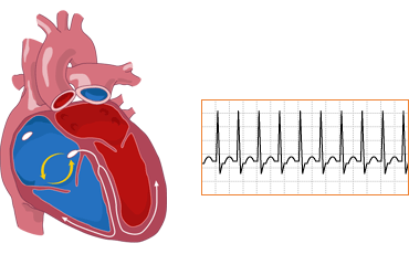
A reentry circuit in the AV node depolarizes the atrium and ventricle simultaneously

ECG Findings:

  • P wave is often buried in QRS or shortly after
  • Rate 150-250 bpm

Treatments:

Multifocal Atrial Tachycardia

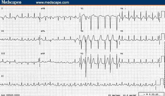
Multifocal Atrial Tachycardia


Etiology:


Signs and Symptoms
Asymptomatic

ECG Findings:
  • Three or more unique P wave morphologies
  • Rate more than 100 bpm
Treatments:
  • Treat the underlying disorder
  • Verapamil or beta blocker